Halal FriendlyTakaful direct billing
YILV CHINA logoYILV CHINA
local_hospitalPartner hospital official material · transcribed verbatimradiology10 radiotherapy and diagnostic units · 13 published casesinfoHospital's own wording · not a promise of outcome

Jinshazhou Hospital of Guangzhou University of Chinese Medicine
Official material — Radiotherapy Oncology Center

This page sets out the radiotherapy platforms, equipment configuration, care pathway and representative cases that the hospital's own promotional material discloses publicly. All copy is transcribed item by item without rewriting; every statement about treatment outcomes is the hospital's own wording and we make no promise on its behalf. Equipment is listed in the order of the hospital's brochure, and cases are grouped by treatment machine with before-and-after imaging.

verified_userUsed with the hospital's permission · patient information de-identifiedlocation_onNo. 1 Lichuandong Street, Jinshazhou, Baiyun District, Guangzhou
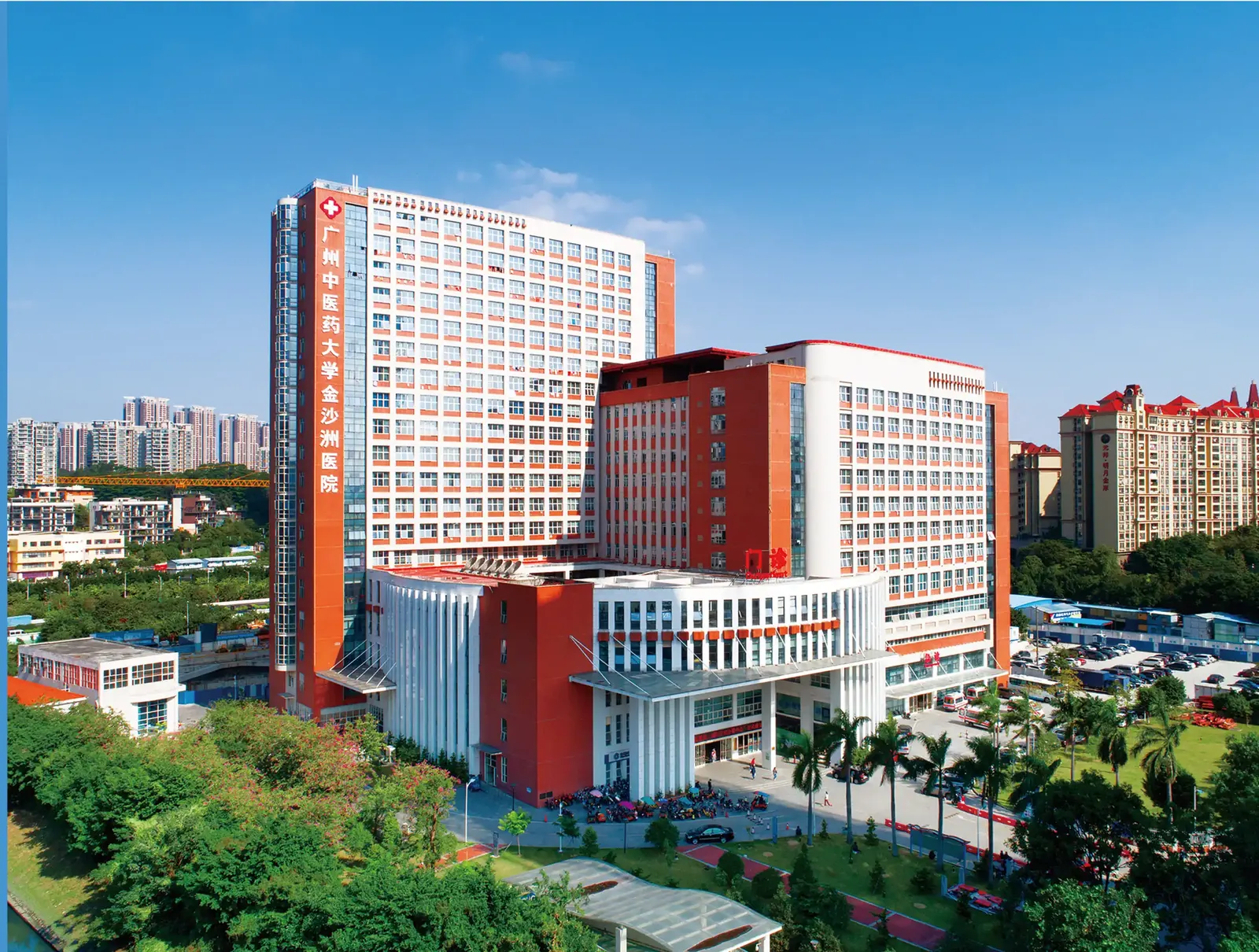
Hospital campus exterior (hospital official material)
SourceOfficial material of Jinshazhou Hospital of Guangzhou University of Chinese Medicine · Radiotherapy Oncology CenterRadiotherapy Oncology Center · Jinshazhou Hospital of Guangzhou University of Chinese Medicine
1,500BedsTertiary Class-III general hospital · approved by the Guangzhou Municipal Health Commission
31Modern laminar flow operating roomsIncluding 5 hundred-level laminar flow operating rooms · 8 interventional operating rooms
48Intensive care (ICU) beds of various typesPlus specialty centers such as blood purification and nuclear medicine
2,113Oncology Medical Center teamProfessional technicians 1,511 · Senior titles 77 / Associate senior titles 103 / Intermediate titles 422
hubFour major platforms

Four major platforms built on equipment

The hospital's installed equipment has reached an internationally leading level and forms four major platforms: diagnostic imaging, tumour radiotherapy, comprehensive tumour treatment and precision testing.

Medical Imaging examination platform

There are over 20 sets of imaging equipment for medical imaging and nuclear medicine imaging. All imaging results can be used for image fusion, truly achieving accurate radiotherapy for multi-modal biological target delineation.

Radiation Oncology platform

Jinshazhou Hospital has established the radiotherapy system in accordance with international standards and installed advanced radiotherapy equipment. Besides, the upcoming proton radiotherapy system has obtained the configuration certificate, and has a complete medical equipment quality control system and information management system.

Tumor comprehensive treatment platform

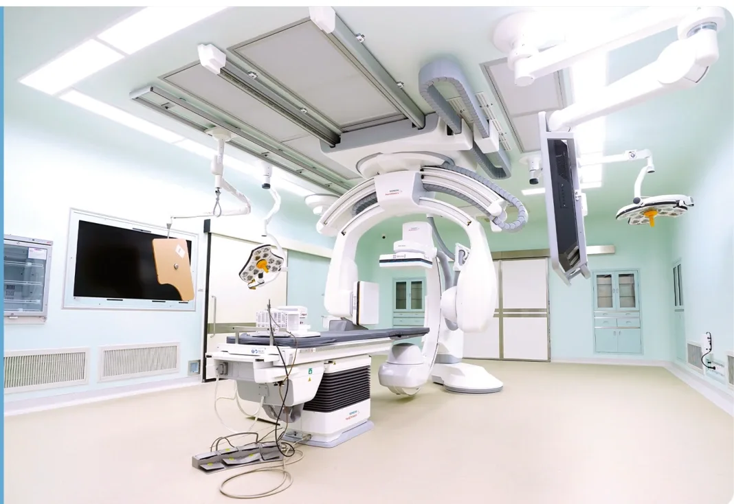
Inclusive of the Da Vinci Robotic Surgery System, DSA Hybrid operating room, blood tumor transplantation cabin, Argon-helium knife cryoablation treatment system, Hifu ultherapy treatment system, acousto-optic Dynamic therapy system, the sixth generation BSD deep tumor phased array hyperthermia system and other equipment.

Accurate inspection platform

Our Biological and Chemical Testing laboratory has reached the ISO15189 standard and the American CAP quality standard. More than 2,600 various inspections can be carried out for companion diagnosis and supplementary diagnosis of tumors.

The centre has a long-term partnership with Australia's ICON Group and has built an international medical team that brings together Chinese and foreign medical specialists. The centre has 2,113 staff, including 1,511 professional and technical personnel, 77 holding full senior professional titles, 103 holding associate senior titles and 422 holding intermediate titles (as at September 2025). The oncology medical centre is built to the standards of China's top-tier (Grade 3A) specialist oncology hospitals and of leading medical institutions, as a modern China–Australia joint oncology treatment centre.

apartmentCampus and facilities

From the campus to the radiopharmacy

Hospital campus exterior (hospital official material)
Campus exterior
Outpatient hall atrium (hospital official material)
Outpatient hall
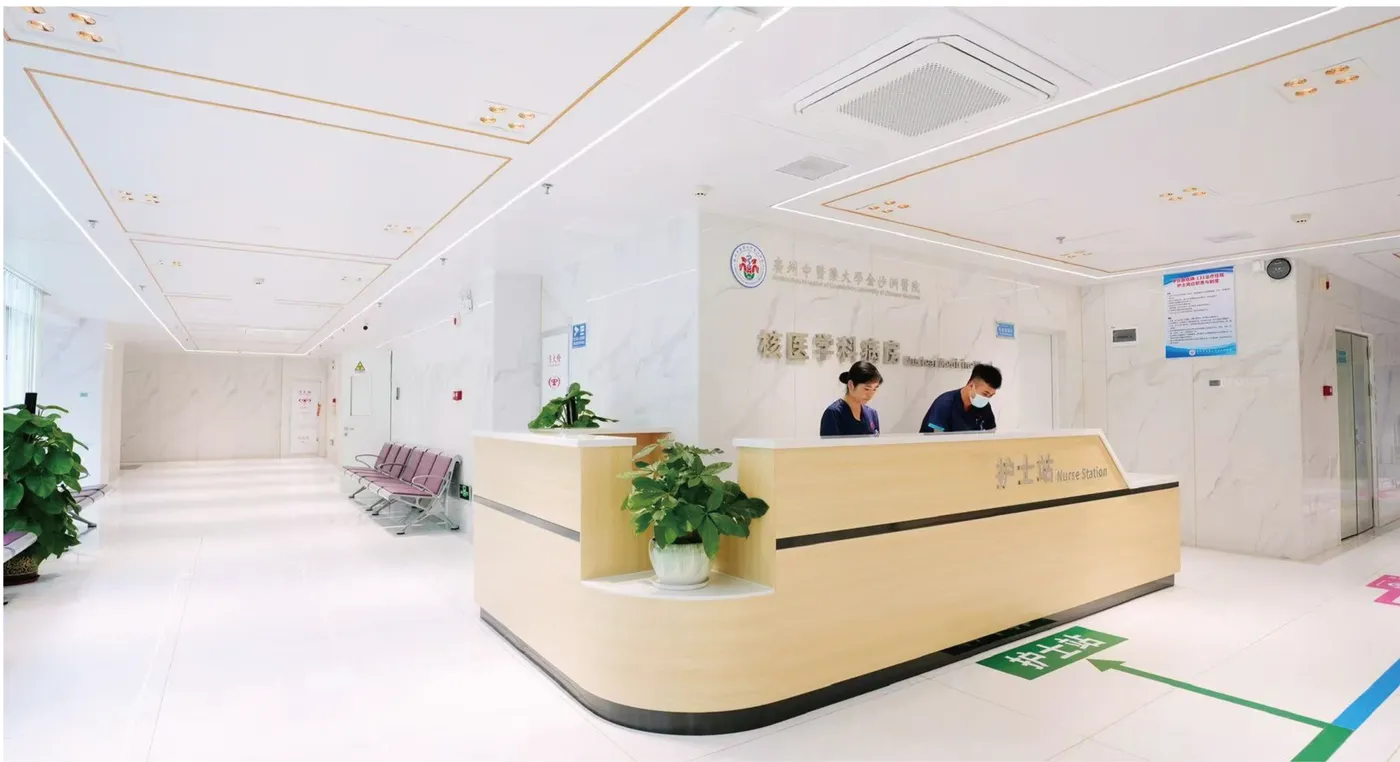
Nuclear medicine reception and waiting area (hospital official material)
Reception and waiting area
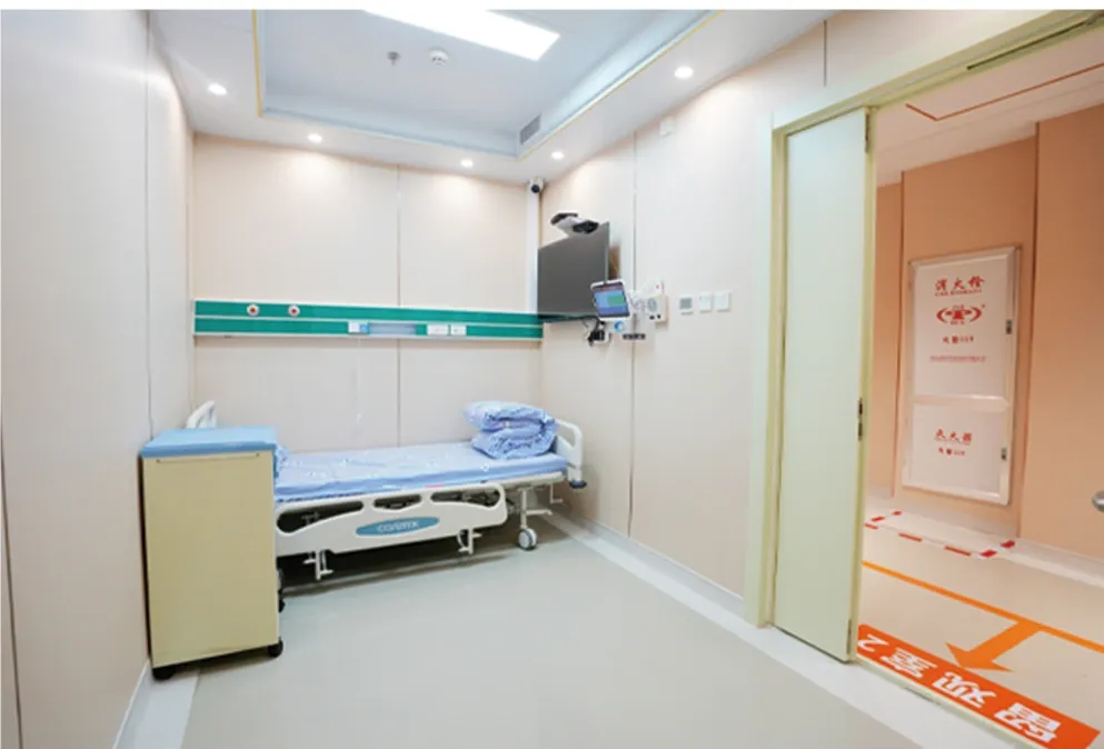
Nuclear medicine ward (hospital official material)
Ward environment
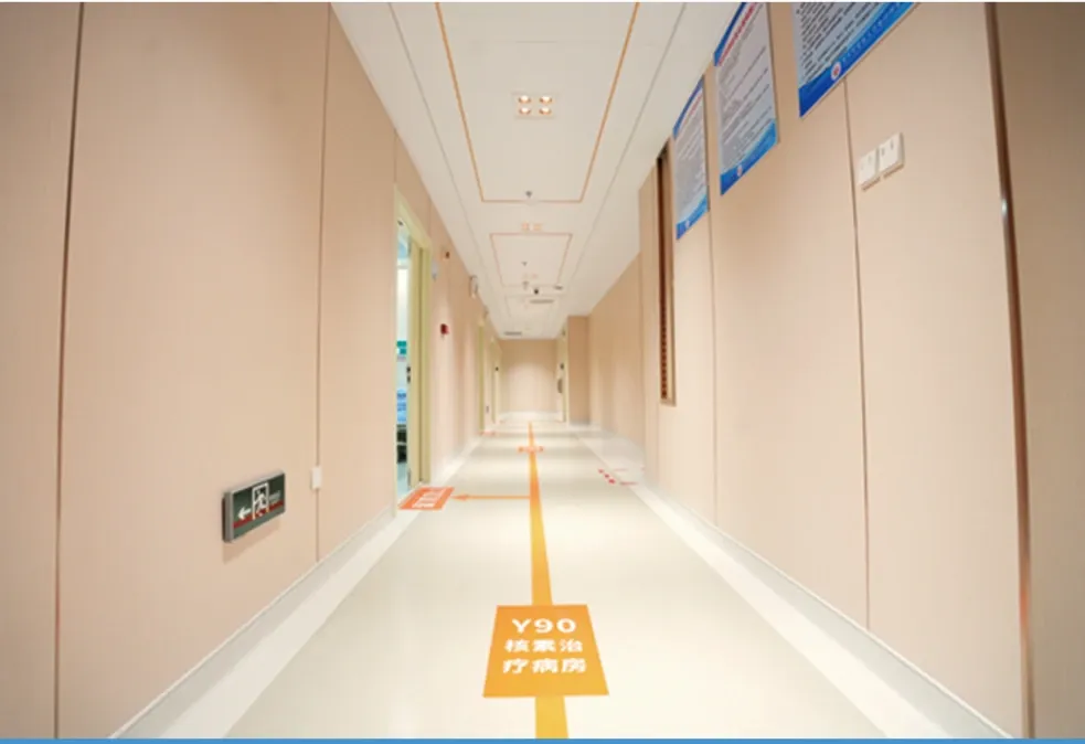
Hospital corridor and wayfinding (hospital official material)
Wayfinding
Cyclotron room at the PET radiopharmacy (hospital official material)
Radiopharmacy
alt_routeMultidisciplinary care and the radiotherapy workflow

Six stages from consultation to follow-up

The multidisciplinary tumour board (MDT) model brings together specialists from surgical oncology, medical oncology, radiotherapy, radiology, pathology and other relevant departments in a working group that proposes the best treatment plan for each patient through regular consultation. The radiotherapy workflow the hospital publishes, together with the estimated time for each stage, is set out below.

1

Consultation with Radiation Oncologist or Inpatient Ward

Patients are consulted by radiation oncology specialists to determine the treatment plan.

2

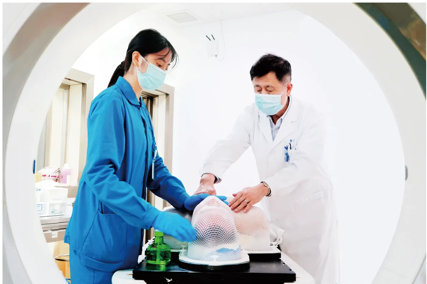
CT Simulation (CT-SIM)

scheduleApproximately 10-30 minutes

Scanning and patient immobilization are completed on the simulation CT as the basis for treatment plan design.

3

Development of Treatment Plan by Radiation Oncologist and Medical Physicist

scheduleApproximately 3-5 working days

Delineate the target volume and organs at risk, and complete dose calculation and plan optimization.

4

Alignment and Verification

scheduleApproximately 10-25 minutes

Reposition according to the treatment plan and verify the position and dose.

5

Treatment

scheduleAccelerator: approximately 5-15 minutes / CyberKnife: approximately 15-60 minutes

Deliver irradiation in fractions according to the plan.

6

Rehabilitation and Follow-up

Post-treatment rehabilitation guidance and regular follow-up examinations.

precision_manufacturingIntelligent precision radiotherapy equipment

10 radiotherapy and simulation units

The equipment below is listed in the order of the hospital's brochure. For each machine, the "What is it", "Indications" and "Advantages" text is the hospital's own wording, transcribed verbatim by this site without verification or endorsement; whether it applies to a patient must be judged independently by the treating physician on the basis of an in-person examination.

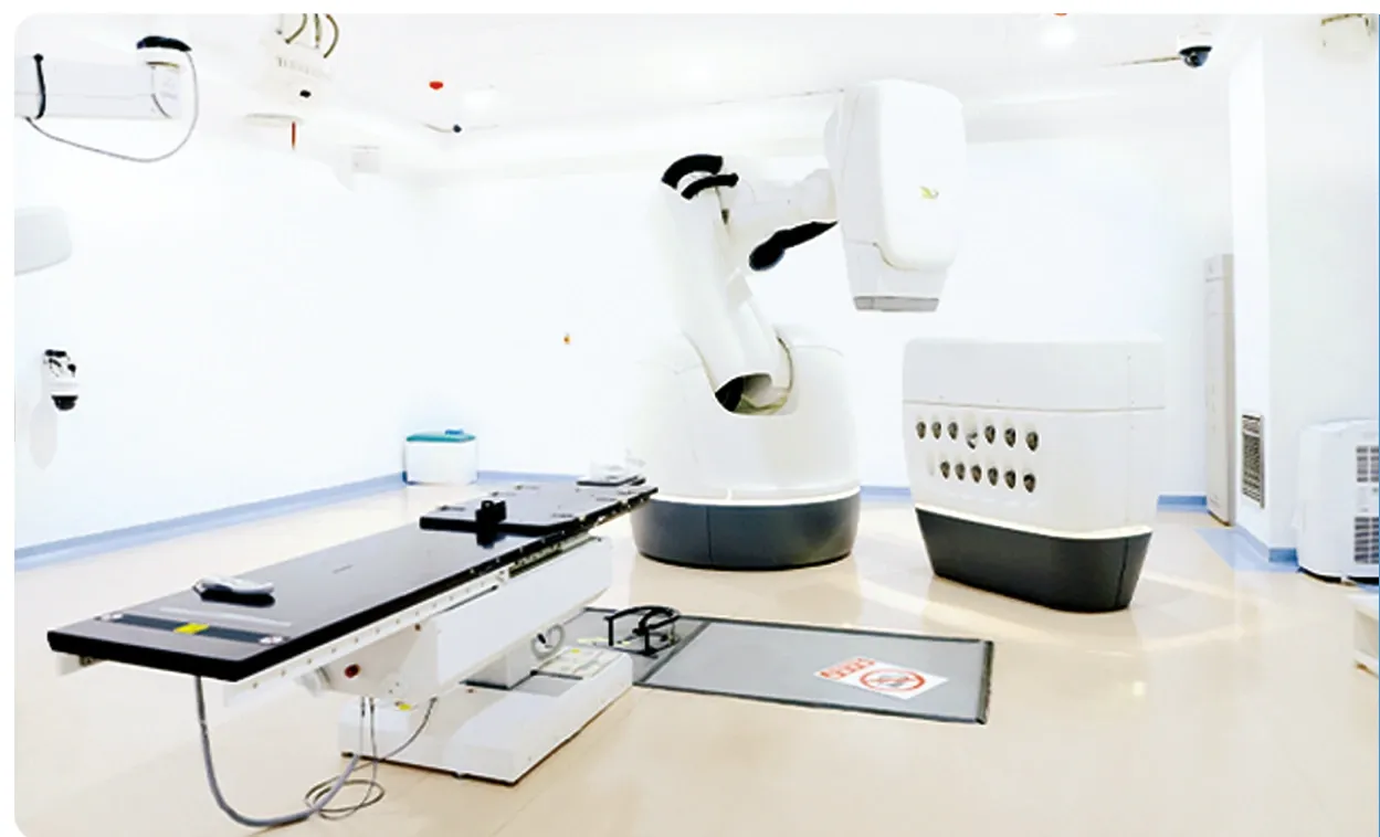
CyberKnife M6 treatment room (official hospital material)

Accuray Sixth-Generation Artificial Intelligence Robotic Radiosurgery CyberKnife

Accuray CyberKnife M6 Robotic Radiosurgery Systemmilitary_techInstalled in 2019 · radiotherapy license obtained on July 7, 2020 · our first case on July 10, 2020
What is Accuray Sixth-Generation Artificial Intelligence Robotic Radiosurgery CyberKnife?

The CyberKnife, also known as the "three-dimensional stereotactic radiosurgery robot". The sixth generation of Accuray CyberKnife (M6) imported by our hospital is a fully digitalized whole-body stereotactic X-ray radiosurgery treatment device with a fully targeted multi-leaf grating. It belongs to the category of stereotactic radiotherapy (SRS/SBRT), which can track the patient's position, tumor location, and patient respiratory motion feedback from three-dimensional directions during treatment. During the treatment process, it can make real-time corrections to the irradiation direction based on medical imaging guidance for the patient's tumor target area (such as tumors).

Advantages
  • checkNon-invasive, painless, no anesthesia, short recovery period, low side effects.
  • checkThe treatment course is short, and only 1-5 times of irradiation are needed to kill the tumor, which can be completed in the outpatient department.
  • checkIt can treat benign and malignant tumors in all parts of the body.
  • checkFast imaging guidance, with unrivaled tumor tracking capabilities.
  • checkThe system error is less than 0.95mm, which means that the tumor irradiation range is smaller and the normal tissue damage is less.
  • checkIt largely eliminates the risk of postoperative complications and significantly improves the quality of life of patients.
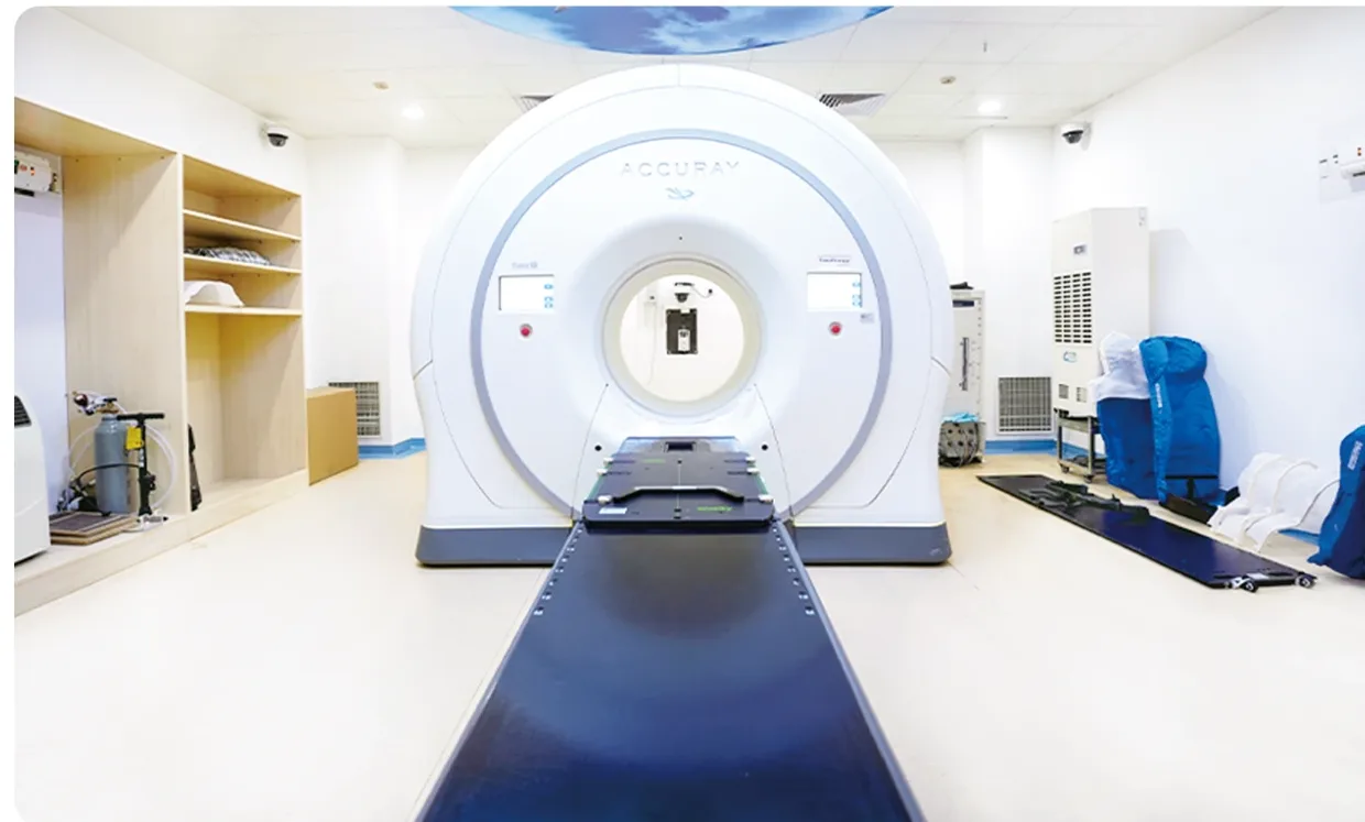
Helical TomoTherapy System treatment room (official hospital material)

Accuray Helical TomoTherapy System

Accuray Helical TomoTherapy Systemmilitary_techIt is a radiotherapy device that utilizes helical CT scanning, the only one of its kind in the world, for treating cancer.
What is Accuray Helical TomoTherapy System?

The TomoTherapy system imported by our hospital integrates IMRT (Intensity-Modulated Radiation Therapy), IGRT (Image-Guided Radiation Therapy), and DGRT (Dose-Guided Radiation Therapy). Its innovative design seamlessly combines a linear accelerator with helical CT, overcoming many limitations of traditional accelerators. With CT guidance, it delivers 360° focal layer irradiation to tumors, enabling efficient and precise treatment for malignant tumor patients.

Indications
  • •The treatment scope of the TOMO system is extensive, applicable to almost all parts of the body, especially complex tumors or those close to vital organs
Advantages
  • checkPrecise Targeting within ±0.1mm: TOMO achieves imaging accuracy of up to ±0.1mm. Its imaging system, comparable to CT, includes dose calculation and verification functions, ensuring dual high precision in patient treatment plans regarding both position and dosage.
  • checkMinimal Side Effects: Clinical summaries from numerous domestic and international hospitals confirm that compared to conventional radiotherapy methods, helical TomoTherapy significantly increases patient survival rates while reducing radiation reactions and complications.
  • checkSimplification through Single-Shot Radiation: TOMO is proficient in tackling complex and challenging tumors, enabling the irradiation of multiple tumor lesions in one session, thus favoring the protection of vital organs.
  • checkWide-ranging Applications: TOMO finds extensive clinical application. It can treat tumors located in any part of the body, covering virtually all cases suitable for radiotherapy, especially those requiring intensity-modulated radiation therapy.
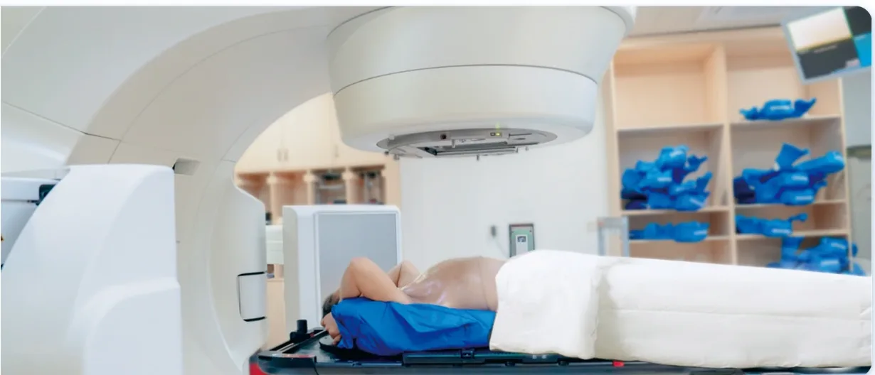
Varian TrueBeam linear accelerator treatment room (official hospital material)

Varian TrueBeam High-Precision Image-Guided Linear Accelerator

Varian TrueBeam Image Guided Linear Acceleratormilitary_techFirst HyperArc case: October 28, 2020 · First respiratory gating case: May 11, 2020
What is Varian TrueBeam High-Precision Image-Guided Linear Accelerator?

The TrueBeam system imported by our hospital is currently the world's most advanced new generation linear accelerator. This system can be used for image-guided radiotherapy and radiosurgery, and is equipped with the new generation of HyperArc technology. Its precise and efficient core features greatly improve the speed and accuracy of tumor treatment, making the device capable of meeting the treatment needs of various radiotherapy techniques and different diseases.

Indications
  • •HyperArc: multiple intracranial brain metastases (fixed-point radiosurgery for several to dozens of lesions can be completed within 10 minutes)
  • •Respiratory gating radiotherapy: lung, liver, breast, abdominal and other tumors that are greatly affected by breathing
Advantages
  • checkIntelligent and reliable quality control system.
  • checkThe most stable and reliable beam characteristics.
  • checkUltra-high-precision mechanical system.
  • checkHigh-definition and high-precision imaging system.
  • checkFeasible stereotactic body radiotherapy (SBRT).
  • checkRespiratory gating radiotherapy technology reduces irradiation of normal tissues and side effects.
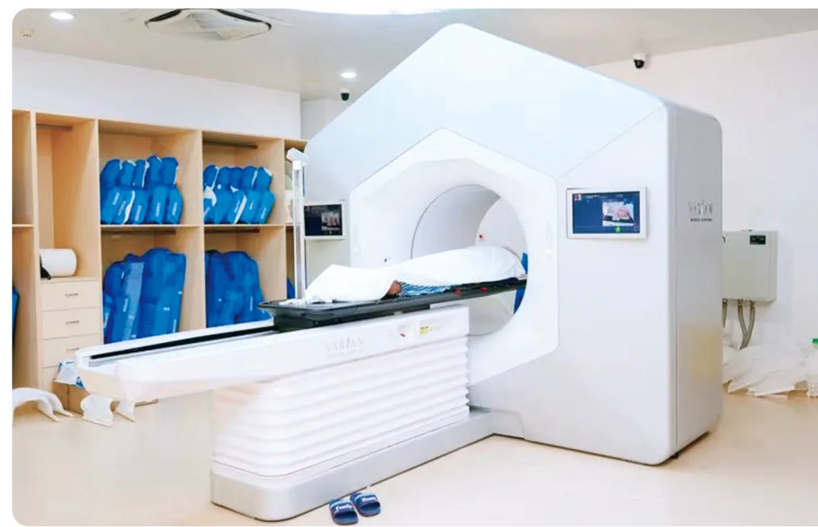
Varian Halcyon Intelligent Linear Accelerator treatment room (official hospital material)

Varian Halcyon Intelligent Linear Accelerator

Varian Halcyon Intelligent Linear Acceleratormilitary_techUpgraded to v4.0 on September 15, 2025, becoming the first device in China equipped with HyperSight image-guided technology
What is Varian Halcyon Intelligent Linear Accelerator?

The Halcyon system imported by our hospital is the world's first medical linear accelerator that achieves 100% image guidance and features full digitalization and artificial intelligence. With the advantages of high integration, high automation, and high intelligence, it is a powerful tool for treating tumors, further elevating radiotherapy from traditional "precision radiotherapy" to "intelligent precision radiotherapy".

Advantages
  • checkHigh speed: 100% coverage of the tumor irradiation area, with the entire treatment process taking only 6-8 minutes.
  • checkHigh precision: the unique "Gemini" dual-layer high-speed multi-leaf collimator system.
  • checkHigh comfort: 100cm large open-bore design, noise-reduction intercom system and water-cooling design reduce patients' fear and irritability.
  • checkHigh intelligence: the blue-ray guidance system simplifies the workflow and greatly reduces manual operation errors.
  • checkHyperSight: the field of view is expanded from 43cm×43cm to 86cm×43cm.
  • checkHyperSight: image quality approaching diagnostic-level and simulation CT quality, directly usable for target delineation and dose optimization.
  • checkHyperSight: iCBCT scan time reduced from 15 seconds to 6 seconds, supporting real-time modification of the target volume and dose recalculation during treatment.
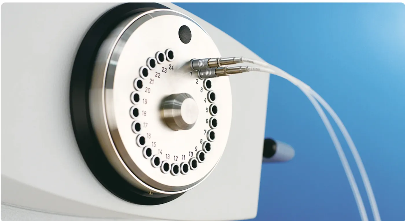
Brachytherapy afterloader radioactive source device (official hospital material)

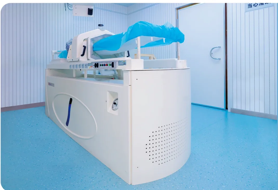
High Dose Rate (HDR) Brachytherapy Afterloader

High Dose Rate (HDR) Brachytherapy Afterloader GammaMedplus iX
What is High Dose Rate (HDR) Brachytherapy Afterloader?

The latest fifth-generation product GammaMedplus™ iX from the American company Varian, introduced by our hospital, is a device specially designed for three-dimensional conformal brachytherapy. Afterloader therapy, also known as brachytherapy, internal radiation therapy, and sealed source radiation therapy, can be a stand-alone treatment or be combined with other treatments such as surgery, external radiation therapy, and chemotherapy to achieve a complementary effect. It uses the natural orifice of the human body (such as the vagina) or inter-tissue insertion to introduce radioactive sources into the body for precise direct irradiation of tumors. It can not only better improve the patient's quality of life and increase the survival rate, but also effectively reduce the risk of local recurrence rate and prolong patient survival.

Indications
  • •Intracavitary treatment: lung cancer, esophageal cancer, rectal cancer, cervical cancer, nasopharyngeal cancer
  • •Interstitial implantation treatment: superficial solid tumors, bone tumors, breast cancer, prostate cancer, tongue cancer
  • •Intraoperative implantation and postoperative treatment: cholangiocarcinoma, pancreatic cancer, liver cancer, abdominal mass
Advantages
  • checkUsing high-activity radioactive sources can achieve high dose rate irradiation of local tumors.
  • checkShort treatment time, and can be used for outpatient treatment.
  • checkHigh safety, effectively reducing side effects and complications, and reducing the local recurrence rate.
  • checkHigh automation, flexible and diverse uterine and vaginal applicators, suitable for different tumor lesions and anatomical structure differences.
  • checkPersonalized treatment plan, obtaining the ideal treatment effect while controlling the dose to adjacent organs.
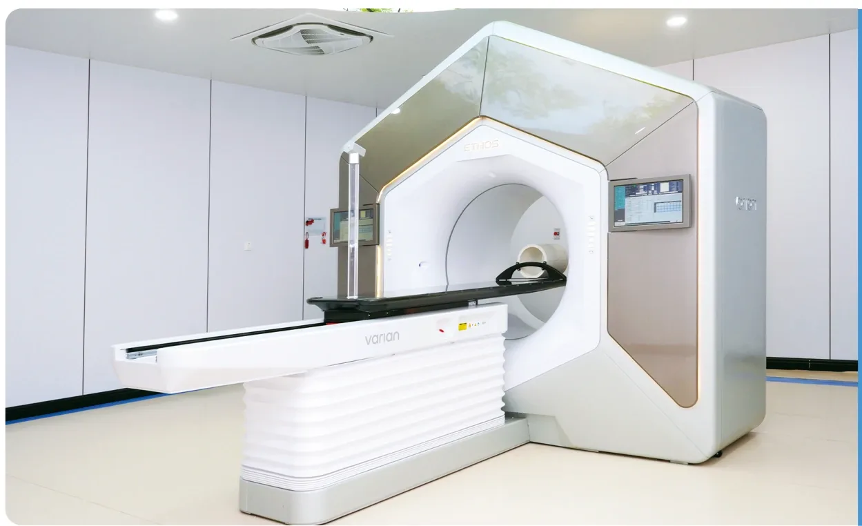
Varian Ethos adaptive radiotherapy treatment room (official hospital material)

Varian Ethos Intelligent Radiotherapy Platform

Varian Ethos Intelligent Radiotherapy Platformmilitary_techInstalled in September 2022 · radiotherapy license obtained on April 7, 2023 · first adaptive radiotherapy case on April 16, 2023
What is Varian Ethos Intelligent Radiotherapy Platform?

The Ethos system imported by our hospital is the world's first online adaptive radiotherapy system based on artificial intelligence technology. It builds upon conventional linear accelerators by incorporating cutting-edge technologies such as artificial intelligence, multimodal high-definition imaging, efficient imaging scanning, and online quality control systems, enabling efficient online adaptive radiotherapy.

Indications
  • •Head and neck tumors, lung cancer, esophageal cancer, breast cancer, prostate cancer and various other common malignant tumors
Advantages
  • checkThe online adaptive radiotherapy of Ethos obtains the patient's current position image before treatment every day, uses artificial intelligence to adjust the target area and modify the plan in real time, so that every time the patient is treated, the radiation can be accurately projected onto the tumor area, achieving accurate individualized radiotherapy every time.
  • checkIt has obvious advantages, especially in prostate cancer, bladder cancer, cervical cancer, endometrioma, rectal cancer and other tumors in parts that change greatly with the filling degree of the intestine and bladder.
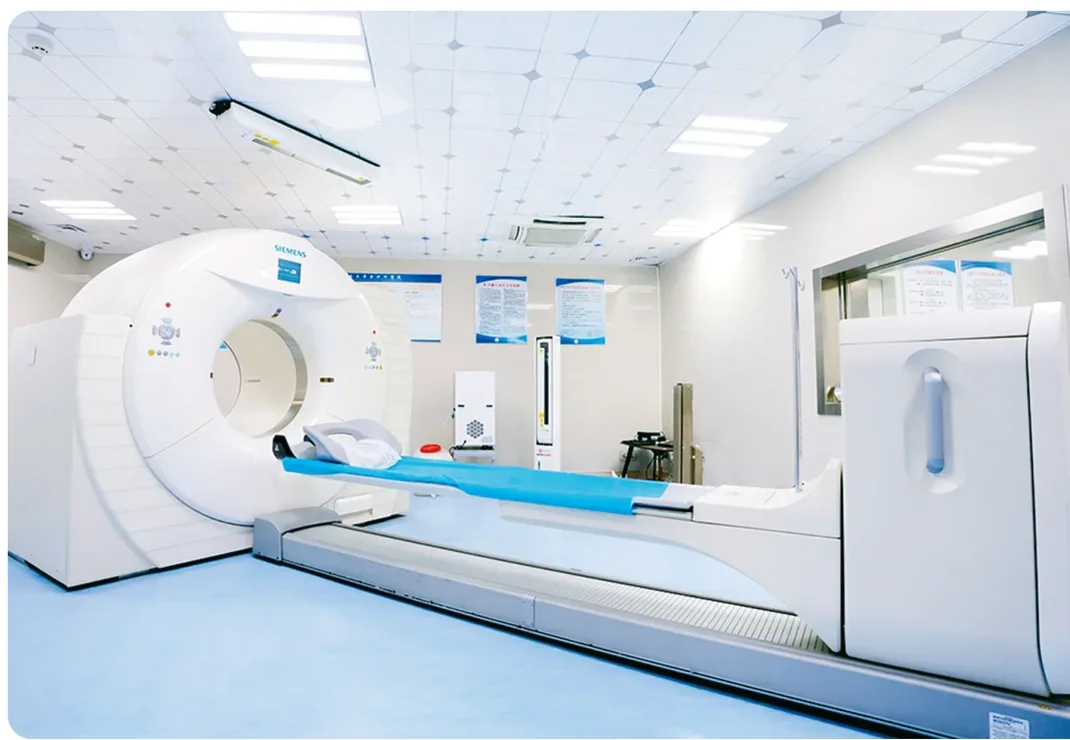
Siemens large-bore simulation CT treatment room (official hospital material)

Siemens New Dual-Energy Four-Dimensional Large-Aperture Positioning Spiral CT

Siemens SOMATOM Confidence Dual-Energy 4D Large-Bore CT Simulator
What is Siemens New Dual-Energy Four-Dimensional Large-Aperture Positioning Spiral CT?

This device achieves the industry's highest multi-layer large-aperture CT, fastest rotation speed, optimal image resolution, and maximum HD FOV (high-definition field of view). In terms of both image quality and accuracy, it enables precise contour delineation and is the basis for precise plan design and treatment, quickly and accurately completing image acquisition for all parts of the body and different positions.

Advantages
  • checkWith its unique dual-energy scanning platform, the equipment not only prevents lesions from escaping detection but also effectively reduces metal artifacts.
  • checkIt is the first positioning CT in the industry capable of achieving "full-aperture" imaging, offering exceptional results for diagnostic examinations of obese patients and specific anatomical regions.
  • checkCapable of conducting self-breath-hold gated imaging scans, this CT simulation positioning device resolves the challenge of treating moving organs during radiotherapy.
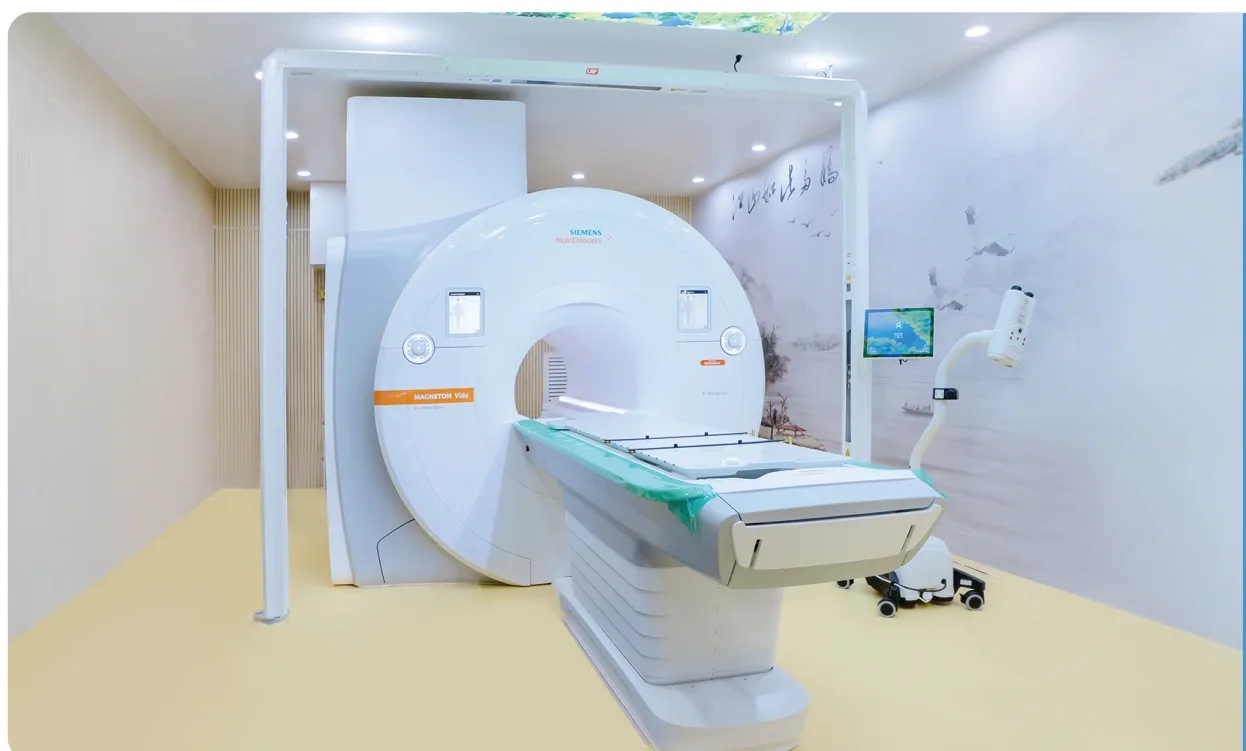
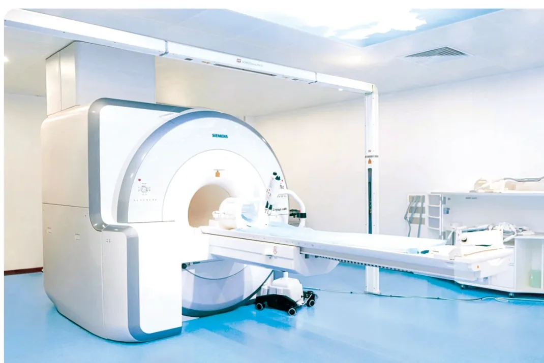
Siemens MAGNETOM Vida simulation MRI treatment room (official hospital material)

Siemens MAGNETOM Vida RT Pro 3.0T Motion-Perception Simulation & Positioning MRI

Siemens MAGNETOM Vida RT Pro 3.0T Motion-Aware MRI Simulator
What is Siemens MAGNETOM Vida RT Pro 3.0T Motion-Perception Simulation & Positioning MRI?

Siemens' MR-only radiotherapy solution enables direct generation of "synthetic CT" images from high-resolution MRI data, completely eliminating radiation exposure. This approach is particularly valuable for pediatric and pregnant patients, and those requiring repeated treatment courses. With a single MRI scan, clinicians achieve both precise target delineation and accurate radiation dose calculation, removing the need for CT simulation and streamlining the entire radiotherapy workflow. This technology is primarily implemented for tumors in the head and pelvic regions.

Advantages
  • checkSelf-gated 4D MRI technology: captures the motion of abdominal and thoracic organs without external gating devices such as respiratory belts.
  • checkPatients can complete radiotherapy simulation and positioning while breathing freely, without breath-holding, improving comfort and removing motion artifacts.
  • checkImproves the positioning accuracy of body tumors, supporting more precise delineation of moving target volumes and organs at risk.
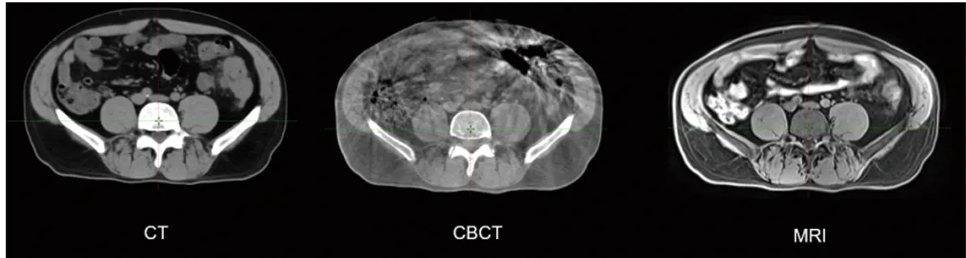
Comparison of CT, CBCT and MRI images (official hospital material)

Distributed MRI Adaptive Technology

Distributed MRI Adaptive Radiotherapymilitary_techOur first case: September 22, 2025
What is Distributed MRI Adaptive Technology?

Prior to treatment, an MRI simulation scan is performed to re-confirm the tumor's position and morphological changes. The radiation oncologist then modifies the target volume accordingly. A rapid planning process is used to re-optimize the treatment plan. Finally, the patient is transferred to the linear accelerator via a shuttle couch, which ensures their position remains unchanged throughout the process. The complete process takes under 25 minutes.

Advantages
  • checkMore Precise Image Guidance: Compared to current CBCT-guided adaptive radiotherapy, MRI provides images free from air-bone artifacts, offers superior soft-tissue resolution, and delivers no additional radiation dose.
  • checkFaster Treatment Delivery: Compared to existing integrated MRI-linear accelerators (MR-linacs), which require approximately one hour for a single online adaptive session, the distributed MRI adaptive radiotherapy system significantly reduces the treatment time. The entire process, including a 6-minute MRI scan and a 6-minute plan optimization, is streamlined to be completed within 30 minutes.
  • checkEnhanced Operational Efficiency: Unlike existing adaptive radiotherapy systems that must pause treatment after imaging to wait for target modification and plan re-optimization, the distributed adaptive approach efficiently coordinates the MRI simulator with the treatment accelerator, eliminating machine idle time.
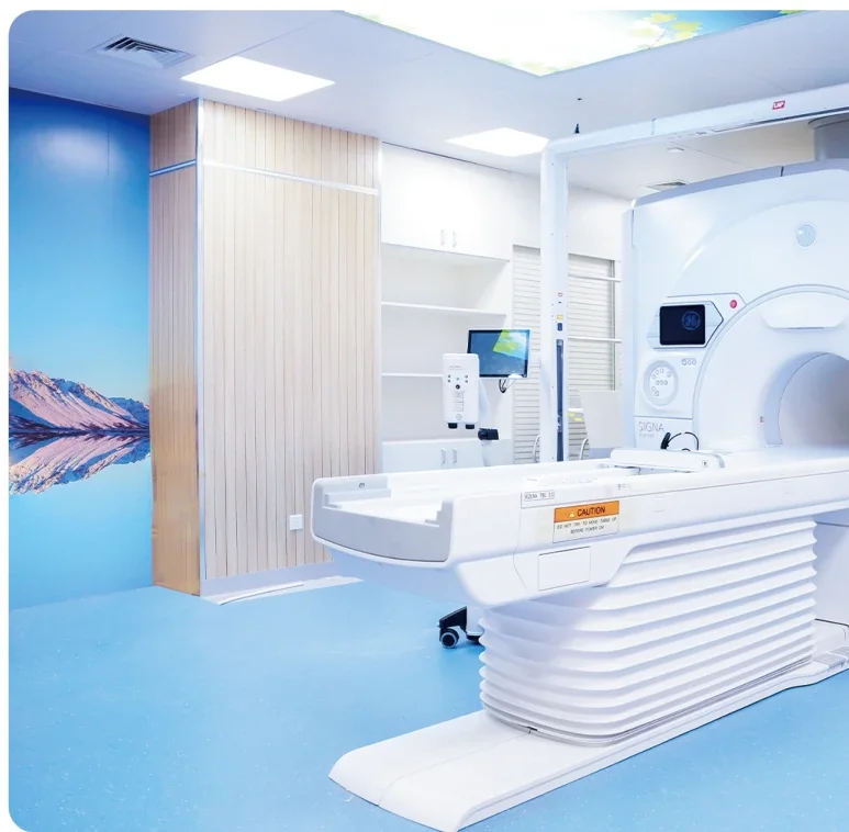
GE SIGNA Premier 3.0T MRI treatment room (official hospital material)

GE SIGNA Premier 3.0T MRI System

GE SIGNA Premier 3.0T MRI System
What is GE SIGNA Premier 3.0T MRI System?

The high-performance, form-fitting exclusive blanket-style AIR Coil delivers unmatched image quality. The Premier model incorporates a new coil material with an INCA fiber loop design, marking a revolutionary breakthrough that makes MRI coils lighter and more flexible. The lightweight, high-channel design of the AIR Coil simplifies radiotherapy patient positioning and significantly improves the visualization of small lesions and tissue boundary sharpness, achieving optimal imaging quality.

Advantages
  • checkIt is the globally unique MRI system capable of achieving an 80 mT/m gradient field strength within a large-bore design. Combined with an internationally leading 148-channel RF platform, it enables high-channel-count imaging for faster scan speeds.
  • checkA complete set of standardized radiotherapy simulation and localization scanning protocols, covering all body regions with standardized sequences and post-processing, delivers superior imaging efficiency.
  • checkA specialized deformation control design with torque-balanced gradient damping combines an industry-best, large-bore magnet with superior field homogeneity and an exclusive 3D gradient deformation correction technology (Gradwrap), effectively overcoming the inherent geometric distortions of MRI and providing highly precise spatial localization for accurate radiotherapy.
scienceNuclear medicine and the PET radiopharmacy

Radionuclide therapy and molecular imaging

The Department of Nuclear Medicine of Jinshazhou Hospital of Guangzhou University of Chinese Medicine boasts a skilled and dedicated team of physicians, nurses, and technologists who are committed to clinical excellence, education, and research. It provides comprehensive nuclear medicine imaging diagnostics and radionuclide therapy. Currently, the department has a total of 37 staff members.

Iodine-131 Therapy for Differentiated Thyroid Cancer (DTC)

Recognized as one of the earliest examples of targeted therapy, molecular imaging, and theranostics, I-131 therapy constitutes an essential element in the triple-therapy regimen for DTC ("surgery + I-131 therapy + TSH suppression"). The clinical portfolio of our department encompasses the application of I-131 SPECT-CT image fusion for diagnosing metastases, as well as molecular targeted therapy for radioiodine-refractory DTC (RAIR-DTC).

Iodine-131 Therapy for Hyperthyroidism

This method is characterized by its simplicity, well-established efficacy, low recurrence rate, minimal side effects, and high safety profile. It is particularly suitable for patients with significantly enlarged thyroids, as well as those for whom antithyroid drug (ATD) therapy has been ineffective, has resulted in relapse, or has caused adverse effects such as leukopenia, liver impairment, or allergic reactions.

Phosphorus-32 (P-32) Patch Therapy

This method is primarily used for the treatment of keloids and infantile hemangiomas. The procedure is characterized by its simplicity, safety, and proven efficacy.

Strontium-89 Therapy for Metastatic Bone Tumors

This treatment is primarily used to alleviate cancerous pain caused by bone metastases from malignant tumors (such as prostate cancer, breast cancer, lung cancer, etc.). Its main advantage is the long duration of pain relief.

Yttrium-90 (Y-90) Resin Microsphere Selective Internal Radiation Therapy (SIRT)

This treatment is primarily indicated for unresectable malignant liver tumors, such as colorectal cancer liver metastases and primary hepatocellular carcinoma. The therapeutic process begins with a pre-treatment planning phase involving nuclear medicine SPECT-CT mapping to assess the patient's suitability for treatment and calculate the therapeutic dosage. Subsequently, the Y-90 resin microspheres are delivered directly to the target tumors via the hepatic artery during an interventional radiology procedure. The tumors are then destroyed by the beta radiation emitted by the Y-90. (The hospital has 4 Y-90 observation wards.)

Diagnostic imaging equipment

Siemens Intevo BOLD SPECT-CTUsed for nuclear medicine imaging diagnostics and pre-treatment Y-90 mapping assessment.
Siemens 52-ring PET/CTTriple breakthrough: ultra-low scan dose, ultra-wide axial field of view (whole-body scanning can be completed in 5 minutes), and ultra-high resolution.
Siemens Fourth-Generation Compass PET/MRI (Biograph Vision Quadra)The ultra-high-sensitivity detector and wide field of view allow a whole-body scan to be completed in as little as 1 minute, with an extremely low radiation dose, making it suitable for children, the elderly and regular follow-ups. Compass technology is based on an MR sequence-based 4D motion model and eliminates respiratory and cardiac motion artifacts without gating.

PET Radiopharmacy Center of the Department of Nuclear Medicine

Cyclotron room of the PET Radiopharmacy Center (official hospital material)

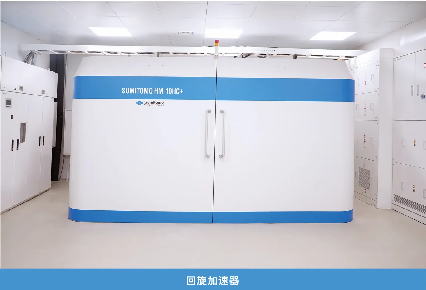
The PET Radiopharmacy Center at the Department of Nuclear Medicine of Jinshazhou Hospital of Guangzhou University of Chinese Medicine is a fully integrated facility for the production and research of positron-emitting radiopharmaceuticals. Operating under full-process Good Manufacturing Practice (GMP) compliance—from its design and construction to daily management—the center is officially licensed by the Guangdong Provincial Medical Products Administration with a "Radioactive Drug Use License (Type 4)" and a "Filing Approval for Positron-emitting Radiopharmaceuticals." The center's infrastructure includes a GMP cleanroom, a cyclotron vault and control room, synthesis hot cells, a quality control (QC) lab, and a small animal lab. Its core equipment is a Sumitomo HM-10HC+ medical cyclotron (10 MeV), equipped with six targets (18F, 11C, 13N, 68Ga, 64Cu, 89Zr), including solid targets for 68Ga, 64Cu, and 89Zr. Currently producing and developing 39 different positron-emitting radiopharmaceuticals, the center serves as a crucial platform for clinical diagnostics and drives the research and clinical translation of novel radiopharmaceutical agents.

biotechTumor Imaging

  • Glucose Imaging: 18F-FDG
  • Tumor Fibroblast Activation Protein Inhibitor Imaging: 18F-FAPI04
  • Prostate Cancer Imaging: 18F-PSMA1007, 68Ga-PSMA11
  • Brain Tumor Imaging: 11C-MET, 18F-FDOPA, 18F-FET, 18F-FLT
  • Breast Cancer Imaging: 18F-FES
  • Liver Tumor Imaging: 18F-FAPI, 11C-Choline
  • Bone Tumor Imaging: 18F-NaF
  • Neuroendocrine Tumor Imaging: 68Ga-DOTATATE, 68Ga-DOTANOC, 68Ga-DOTATOC, 18F-Octreotide, 18F-FDOPA
  • Pheochromocytoma Imaging: 18F-mFBG, 18F-FDOPA
  • Parathyroid Imaging: 11C-Choline
  • Tumor Hypoxia Imaging: 18F-FMISO

biotechNeuropsychiatric Imaging

  • Neuroinflammation TSPO Imaging: 18F-DPA714, 18F-FEPPA
  • Cerebral Blood Flow Imaging: 13N-Ammonia
  • AD Brain Tau Protein Imaging: 18F-AV1451
  • AD Brain Aβ Amyloid Protein Imaging: 11C-PIB, 18F-AV45
  • Epileptogenic Focus Imaging: 18F-FDG, 18F-FMZ, 18F-UCB-H
  • Brain Dopamine Imaging: 18F-FDOPA
  • PD Brain Dopamine Transporter (DAT) Imaging: 11C-CFT
  • PD Cardiac Sympathetic Nerve Imaging: 18F-mFBG

biotechCardiac Imaging

  • Myocardial Blood Flow Perfusion Imaging: 13N-ammonia
  • Myocardial Viability Imaging: 13N-ammonia + 18F-FDG
  • Myocardial Fibrosis Imaging: 13N-ammonia + 18F-FAPI
  • Cardiac Sympathetic Nerve Imaging: 18F-mFBG
devices_otherOther equipment

Comprehensive treatment and imaging equipment

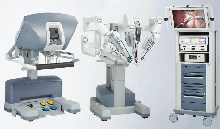
Da Vinci Surgical RobotDa Vinci Surgical Robot
Siemens Artis icono biplaneSiemens Artis icono biplane
Siemens PET-CT Biograph mMR with COMPASSSiemens PET-CT Biograph mMR with COMPASS
Siemens 3.0T 64-ring PET-MR Biograph mCTSiemens 3.0T 64-ring PET-MR Biograph mCT
BSD-2000 Hyperthermia MachineBSD-2000 Hyperthermia Machine
FreeGS-A Gamma KnifeFreeGS-A Gamma Knife
Da Vinci surgical robot (hospital official material)
DSA hybrid operating theatre (hospital official material)
Deep tumour hyperthermia equipment (hospital official material)
PET-CT suite (hospital official material)
Diagnostic imaging suite (hospital official material)
clinical_notesRepresentative cases in intelligent precision radiotherapy

Published cases grouped by treatment machine

13 cases are grouped by treatment machine, with before-and-after imaging published by the hospital. All diagnoses, doses and follow-up results are transcribed from the hospital's own text; an individual result does not mean that similar conditions will all achieve the same result. Imaging is taken from the hospital's public material, Used with the hospital's permission · patient information de-identified.

CyberKnife (M6) Treatment Cases

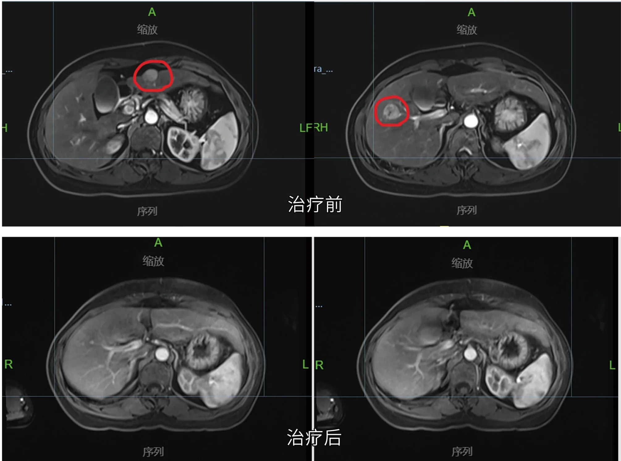
Pre- and post-treatment MR comparison of a liver cancer patient treated with CyberKnife (official hospital material)
CyberKnife3 cases
  1. 10-year-old female · Brain Malignant Tumor (Glioma)Treatment: Started CyberKnife radiotherapy on June 14, 2020; radiation dose to PTV: 25 Gy in 5 fractions (5 Gy per fraction)Follow-up: Follow-up imaging two months later showed a slight reduction in the brainstem tumor. The patient regained consciousness, with significant improvement in speech function and gradual normalization of limb movements.
  2. 67-year-old male · Secondary Malignant Brain TumorTreatment: Initiated CyberKnife radiotherapy on August 3, 2020; radiation dose to 95% GTV: 15 Gy in 3 fractions (5 Gy per fraction)Follow-up: Imaging review three months later shows a significant reduction in the tumor size compared to previous scans.
  3. 46-year-old female · Liver CancerTreatment: Treatment plan involved 4D simulation positioning, with tumors in segments S2 and S3 receiving a dose of 50 Gy in 5 fractions (10 Gy per fraction)Follow-up: December 6, 2020: Pre-treatment MR examination showed multiple nodules in liver segments S2, S3, and S5, consistent with likely hepatocellular carcinoma. April 14, 2021: Follow-up MR imaging indicated a significant reduction in the liver cancer lesions compared to previous scans.

TOMO and TrueBeam Treatment Cases

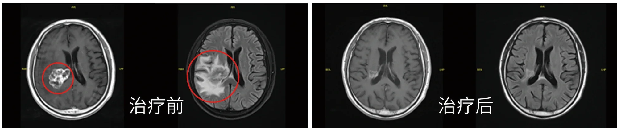
Pre- and post-radiotherapy imaging comparison of an intracranial tumor (official hospital material)
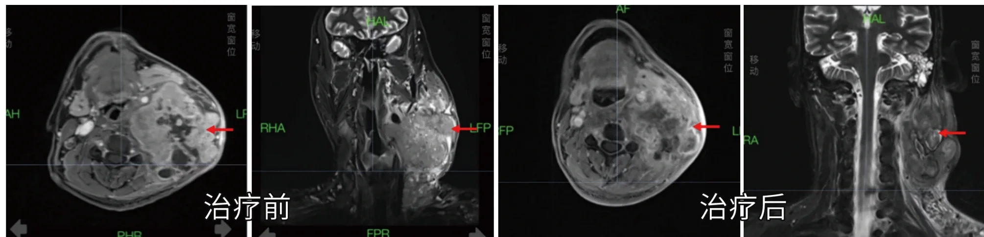
Pre- and post-radiotherapy imaging comparison of a head and neck tumor (official hospital material)
TOMOTrueBeam4 cases
  1. Female, 16 years old · Malignant Brain Tumor (Intracranial Germ Cell Tumor, WHO Grade IV)Treatment: Started TOMO radiotherapy on May 19, 2022; radiation dose to PTV: 45 Gy in 25 fractions (1.8 Gy per fraction)Follow-up: Follow-up imaging 10 months later showed a significant reduction in tumor size in the saddle and pineal regions compared to previous scans.
  2. Male, 47 years old · Nasopharyngeal Malignant Tumor (Undifferentiated Non-keratinizing Carcinoma, cT4N3M1, Stage IV)Treatment: Started TOMO radiotherapy on November 30, 2022; PGTV: 69.96 Gy in 33 fractions (2.12 Gy per fraction), PTVn: 66 Gy in 33 fractions, PTV: 60 Gy in 33 fractionsFollow-up: Three months after treatment, imaging revealed a significant reduction in the size and number of metastatic lymph nodes in the neck compared to previous scans.
  3. Male, 66 years old · Nasopharyngeal malignant tumor (Undifferentiated non-keratinizing carcinoma of nasopharynx)Treatment: April 12, 2021, to May 27, 2021: VMAT radiotherapy with a prescribed dose to the planning gross tumor volume of 69.96 Gy in 33 fractions (2.12 Gy per fraction)Follow-up: After-treatment continuous follow-up examinations revealed gradual shrinkage of the nasopharyngeal mass; MRI results on January 5, 2023, showed complete disappearance of the nasopharyngeal mass, with a significant reduction in previously positive lymph nodes.
  4. Male, elderly · Lung cancer with brain metastases, accompanied by brain dysfunction, requiring wheelchairTreatment: 5 sessions of radiotherapyFollow-up: Before treatment: severe peritumoral edema with elevated intracranial pressure. After 5 sessions of radiotherapy, tumor regression was observed; brain function recovered and the patient regained the ability to walk independently.

Halcyon and Brachytherapy Afterloader Treatment Cases

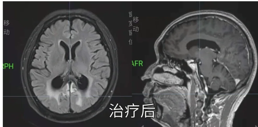
Post-radiotherapy imaging of intracranial lymphoma (official hospital material)
Pre-brachytherapy MR imaging of cervical cancer (official hospital material)
HalcyonBrachytherapy Afterloader4 cases
  1. 49-year-old male · Diffuse Large B-cell LymphomaTreatment: March 11, 2021: Radiation therapy for intracranial lesions using 6MV-X-rays. Synchronous boost irradiation with a dose of 40 Gy in 20 fractions to 95% of PTV1, and 36 Gy in 20 fractions to PTV2, administered 5 times per weekFollow-up: April 7, 2021: Follow-up MRI showed a significant reduction in intracranial lesions, indicating a positive response to lymphoma treatment.
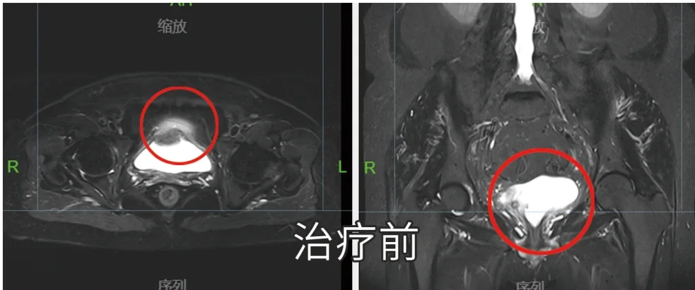
  2. 59-year-old female · Bladder Cancer (March 29, 2021 PET/CT revealed multiple lesions in the anterior, posterior, and bottom walls of the bladder, suggestive of tumor recurrence)Treatment: Synchronous chemoradiotherapy (single-agent cisplatin 20 mg/m² weekly); pelvic VMAT 45 Gy, with a local boost of 20 Gy to the bladder, using image-guided radiation therapy (IGRT)Follow-up: Post-radiotherapy pelvic MRI one month later showed no residual tumor or signs of recurrence in the bladder.
  3. 53-year-old female · Diagnosed Stage IB Cervical Cancer in February 2020Treatment: The patient had no surgical indication and underwent chemotherapy at another hospital with unsatisfactory results; definitive radiotherapy for cervical cancer at our hospital (external beam radiotherapy + brachytherapy)Follow-up: Significant reduction in the size of the tumor observed post-treatment, with complete disappearance of the large tumor.
  4. 73-year-old female · Diagnosed Stage IIIA Cervical Cancer in February 2020Treatment: The patient had no surgical indication and underwent chemotherapy and interventional therapy at another hospital with unsatisfactory results; definitive radiotherapy for cervical cancer at our hospital (external beam radiotherapy + brachytherapy)Follow-up: Substantial reduction in tumor size noted post-treatment, with complete disappearance of the large tumor.

ETHOS Treatment Cases

Target delineation imaging during Ethos adaptive radiotherapy (official hospital material)
Pre-treatment MR imaging (official hospital material)
Ethos2 cases
  1. Male, 55 years old · Nasopharyngeal Malignant Tumor (Undifferentiated Non-keratinizing Carcinoma)Treatment: Prescribed dose: PGTVnx 70.29 Gy in 33 fractions (2.13 Gy per fraction), PGTVnd 67.98 Gy in 33 fractions (2.06 Gy per fraction), PTV1 62.04 Gy in 33 fractions (1.88 Gy per fraction), PTV2 54.12 Gy in 33 fractions (1.64 Gy per fraction)Follow-up: Eight months after treatment, follow-up MR imaging indicated a reduction in the size of the nasopharyngeal mass and positive lymph nodes compared to previous scans. Improvement in nasopharyngeal mucosal thickening was also observed.
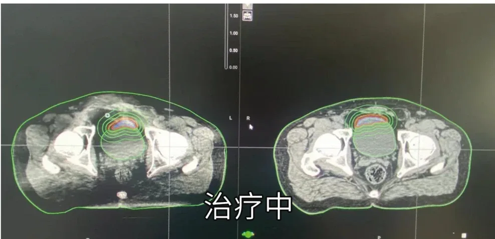
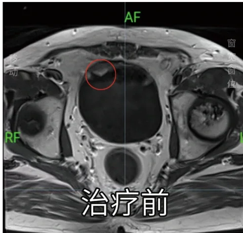
  2. Male, 67 years old · Bladder CancerTreatment: Treatment challenge: tumor position varies with bladder filling, poor reproducibility. ETHOS online adaptive treatment: utilizes iCBCT imaging before each treatment to capture the real-time position of the patient's bladder and tumor, then recontours the target area and redesigns the treatment planFollow-up: Illustrations include pre-treatment MR, target delineation during Ethos adaptive radiotherapy, and post-treatment MR.

The content and imagery above are taken from Official material of Jinshazhou Hospital of Guangzhou University of Chinese Medicine · Radiotherapy Oncology Center and are material the hospital has publicly disclosed. Terms such as "first case", "complete disappearance" and "marked shrinkage" are the hospital's own wording; this site transcribes them verbatim without verification or endorsement. The procedure, dose and outcome of an individual case all depend on stage, pathological type, underlying disease and response to treatment, and none of this constitutes medical advice, a promise of efficacy or a guarantee of future results. Any diagnostic or treatment decision must be made independently by a qualified medical institution and a licensed physician on the basis of the patient's in-person examination results.

support_agentFree pre-assessment · we do not push unnecessary cross-border care

Want to confirm whether your condition matches the radiotherapy equipment and disease areas above?

Submit the diagnostic, pathology and imaging documents you already have from a Malaysian hospital and we will help check the disease area, equipment suitability and the receiving hospital's arrangements. The platform provides standardised record preparation, bilingual medical translation and coordination with Chinese hospitals; the treatment plan is determined independently by the treating physician team at the receiving hospital.